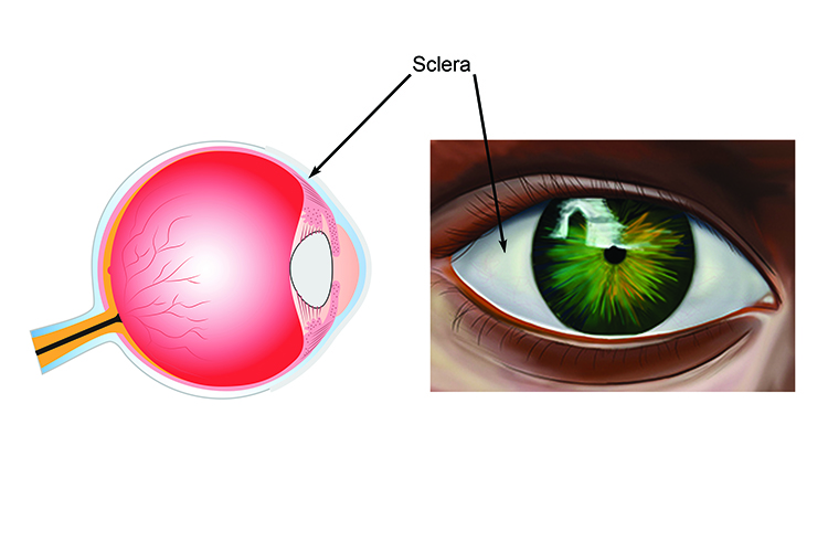
The sclera, the white is the protective wall of the eye ball
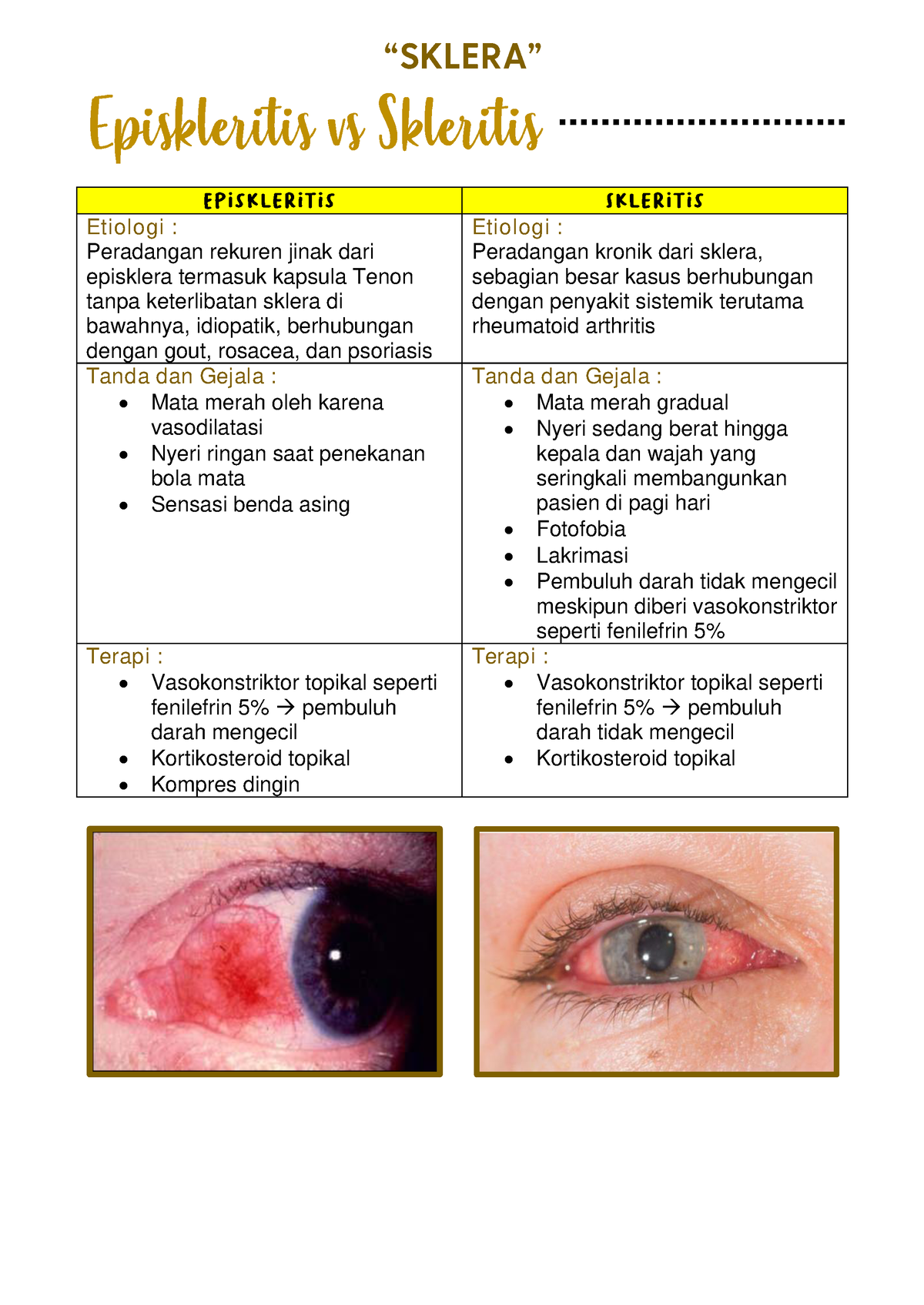
Episcleritis is an inflammation of the outermost layer of the sclera. Symptoms include: Redness. Mild pain. Swelling of the conjunctiva. Eyelid swelling. Raised nodules on the eye. Most cases are sectoral, meaning only a section of the sclera is affected. In diffuse cases, the entire sclera can be affected.
Sklera (Episkleritis dan Skleritis), Kornea, dan Bilik Mata Depan Etiologi Peradangan
Sklera yang keruh dapat dicegah dengan menghindari stres berlebihan, tidak mengonsumsi alkohol, tidak merokok, memiliki jam tidur yang cukup, tidak terpapar matahari terlalu lama, tidak terpapar cahaya gadget terlalu lama, serta banyak mengonsumsi makanan bergizi. Pewarnaan fokal kekuningan pada sklera juga mungkin disebabkan oleh degenerasi.
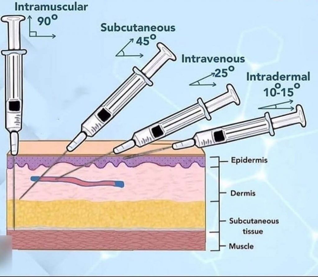
4 Cara Pemberian Obat Injeksi atau Parenteral
7. Glaukoma (peningkatan tekanan bola mata) 8. Skleritis (inflamasi sklera) 9. Trauma bahan kimia. Penyebab kondisi mata merah dapat diketahui melalui wawancara lebih dalam mengenai gejala utama dan tambahan (adakah gangguan penglihatan, adakah kotoran mata, silau, berair, rasa mengganjal, dan lain sebagainya), serta pemeriksaan mata secara.

Lederhaut (Sklera) Anatomie und Funktion MediKarriere
Scleritis is the inflammation in the episcleral and scleral tissues with injection in both superficial and deep episcleral vessels. It may involve the cornea, adjacent episclera and the uvea and thus can be vision-threatening. Scleritis is often associated with an underlying systemic disease in up to 50% of patients.
Mengenal Fungsi Sklera, Bagian Putih pada Bola Mata Hello Sehat
alienum sklera okuli sinistra. Penatalaksanaan non-medikamentosa yaitu tatalaksana operatif berupa ekstraksi corpus alienum sklera, dan penatalaksanaan medikamentosa preoperatif berupa injeksi ceftriaxone 1 gr/12 jam (IV), ketorolac Tromethamine 1ampul/12 jam (IV drip), Injeksi Anti Tetanus Serum (IM),

Tutorial injeksi subkutan 2 YouTube
The Sclera. The sclera is tough and fibrous, protecting the interior components of the eye from injury, and makes up the exterior coating of the eye. The sclera forms the entire visible white exterior of the eye, while the iris is the colored portion inside the anterior chamber of the eye. Although we can only see the visible portion of the.

Pin on EENT
Pleksus vaskular dalam terletak di lapisan permukaan sklera, dan stasis maksimum yang terkait dengan sklerit dikaitkan dengan hal itu. Pada saat yang sama, beberapa injeksi bejana permukaan tidak bisa dihindari, tapi ini tidak signifikan. Pembesaran fenilefrin tidak mempengaruhi pembuluh melebar dari pleksus ini.

Bab 07. Mata Merah haryahutamas Halaman 2 PDF Online PubHTML5
Pada mata normal sklera terlihat berwarna putih karena sklera dapat terlihat melalui bagian konjungtiva dan kapsul tenon yang tipis dan tembus sinar.. - Pupil ukuran normal dengan reaksi normal. Injeksi Siliar Melebarnya pembuluh darah perikornea (a. Siliar anterior) atau injeksi siliar atau injeksi perikornea terjadi akibat radang kornea.

INJEKSI IV, IM, IC, SC. YouTube
Ketika pembuluh darah di bawah konjungtiva pecah, darah akan memenuhi area di antara konjungtiva dan sklera. Akibatnya, bagian putih mata akan terlihat bercak berwarna kemerahan. Penyebab Perdarahan Subkonjungtiva. Penyebab perdarahan subkonjungtiva tidak selalu diketahui secara pasti. Namun, pembuluh darah pada bagian ini memang cenderung.
Arrow showing connective tissue bands between sclera and Tenon capsule... Download Scientific
injeksi sklera di superior, bayangan koroid di superonasal, perdarahan subkonjungtiva, bleb terbentuk, hekting intak, mikrokornea, sklerokornea jam 12 sampai 3, kekeruhan di perifer 360 derajat, dan edema kornea. BMD Van Herrick grade I-II f/c sulit dinilai, pupil irregular, iris transillumination defect (+), sinekia
:max_bytes(150000):strip_icc()/GettyImages-87299101-59a0952c396e5a0011d753d6.jpg)
Fungsi Sklera pada Mata Manusia Dewan Medis
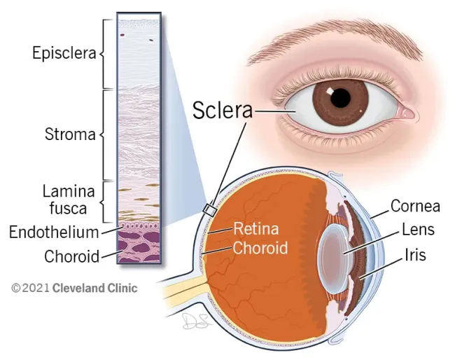
The sclera is the opaque white posterior part of this outer layer. It protects the inner eye contents and gives structural integrity to the eye. The sclera is continuous anteriorly with the cornea and posteriorly with the dural sheath of the optic nerve. Learn everything about the anatomy of the eyeball with the following study unit!

Kuliah Sklera PDF
Sklera melapisi bola mata dari ujung kornea hingga ke saraf optik di belakang mata. Berdasarkan bagian sklera yang mengalami peradangan, skleritis dibagi menjadi dua, yaitu skleritis anterior (sisi depan bola mata) dan skleritis posterior (sisi belakang bola mata). Skleritis dapat terjadi hanya pada satu bagian mata, namun ada juga yang terjadi.
Sclera Aufbau, Funktion & Krankheiten MedLexi.de
1. INTRODUCTION. Ocular complaints are responsible for approximately 2-3% of patient visits to primary care physicians and emergency facilities, of which the majority are for the management of conjunctival hyperemia.[1-3] However, the current literature reports limited evidence about the prevalence of conjunctival hyperemia due to its self-limiting nature.

Case Ruptur Sklera & Hifema
Scleritis is a severe ocular inflammatory condition affecting the sclera, the outer covering of the eye. It can be categorized as anterior with diffuse, nodular, or necrotizing subtypes and posterior with diffuse or nodular subtypes. Scleritis can be visually significant, depending on the severity and presentation and any associated systemic conditions.[1] The presentation can be unilateral or.

Anatomi Sklera PDF
The sclera, also known as the white of the eye or, in older literature, as the tunica albuginea oculi, is the opaque, fibrous, protective outer layer of the eye containing mainly collagen and some crucial elastic fiber.. In the development of the embryo, the sclera is derived from the neural crest. In children, it is thinner and shows some of the underlying pigment, appearing slightly blue.

SOP Lengkap Injeksi Intra Muskuler (IM) Di Kelas Teori & Praktek STIKESMUS YouTube
Melansir My Cleveland Clinic, fungsi sklera antara lain: menjaga bentuk bola mata, dan. melindungi bola mata dari cedera. Otot-otot yang menempel pada sklera mata bertugas untuk menggerakkan mata ke atas, ke bawah, dan ke samping. Keseluruhan sklera dan kornea mata kemudian dilapisi oleh membran sangat tipis dan jernih yang disebut konjungtiva.