
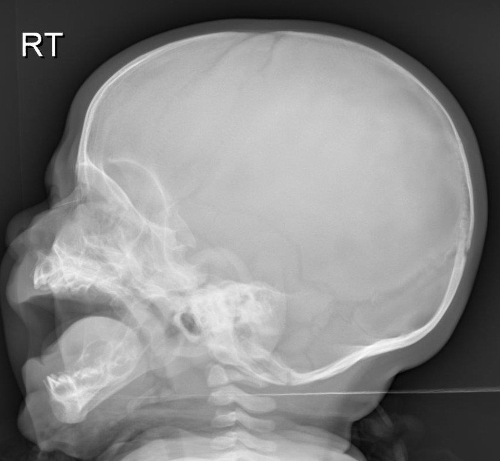
Normal Skull XRay Lateral View
1. Normal Skull Anatomy. Recognizing Symmetry: Normal Cranial Features showcase a balanced appearance of cranial bones. Clear Delineation: The skull base and vault should be distinctly outlined. Expected Contours: Familiarize with the typical contours of facial bones. 2. Fractures and Trauma Indicators. Cracks and Signs of Impact: Identify linear or depressed fractures and signs of impact.

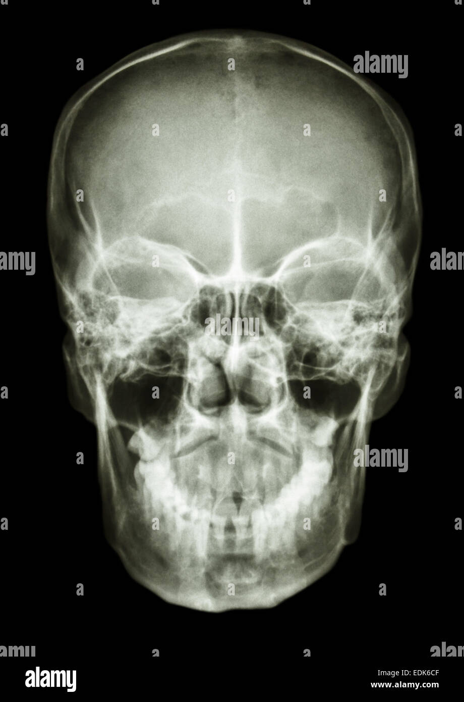
Ap Skull X Ray Normal skull xray Image / Ap
Introduction The skull is a compact structure that covers and protects the brain and facial organs. [1] [2] It is in a complex anatomical relationship with many craniofacial organs and associated tissues, each of which has a different embryological origin and performs different functions.

Skull X Ray Anatomy anatomy diagram source
The skull is the skeletal framework of the head of vertebrates formed by the cranium. It encompasses the frontal, parietal, occipital, temporal, zygomatic, lacrimal, nasal, ethmoid, and sphenoid bone as well as the maxilla. This chapter illustrates the normal CT anatomy of the skull. Keywords Jaw Normal anatomy Download reference work entry PDF

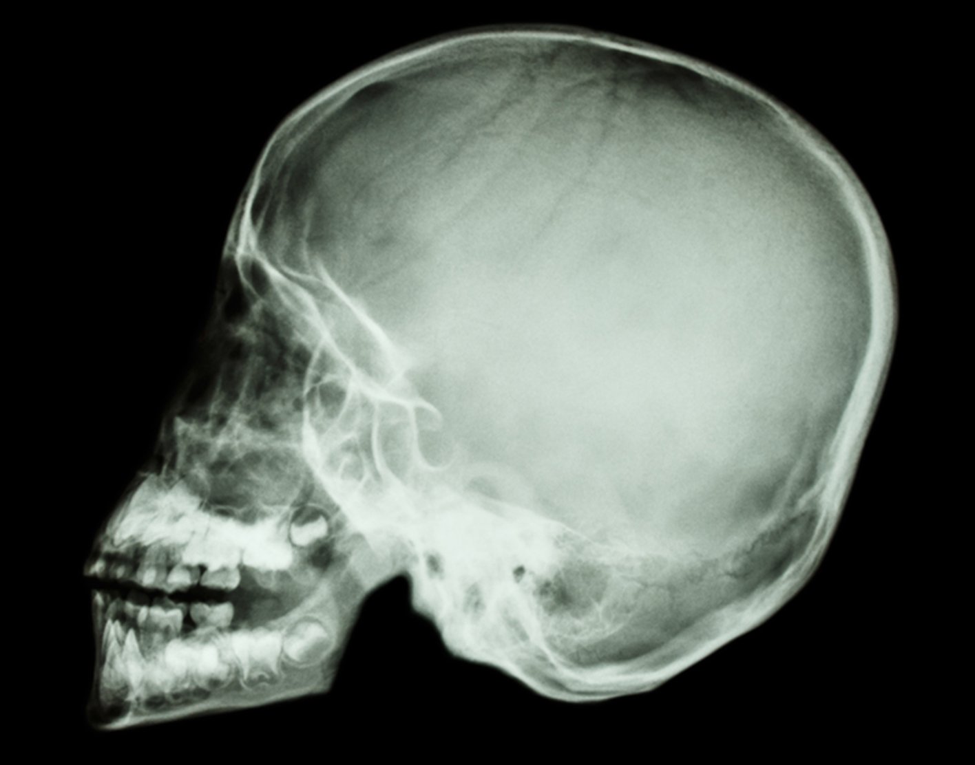
Lateral Xray of Skull Stock Image C043/0235 Science Photo Library
What is a skull X-ray? X-rays use invisible electromagnetic energy beams to make images of the skull. Standard X-rays are done for many reasons, including diagnosing tumors, infection, foreign bodies, or bone injuries. X-rays use external radiation to produce images of the body, its organs, and other internal structures to diagnose a problem.
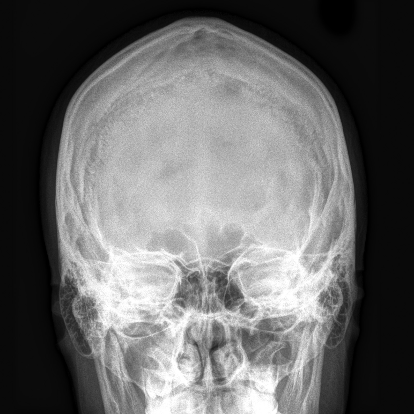
film xray skull AP show normal human's skull Stock Photo 77254143
An X-ray of the skull follows this general process: You will be asked to remove any clothing, jewelry, hairpins, eyeglasses, hearing aids, or other metal objects that might interfere with the X-ray. If you are asked to remove clothing, you will be given a medical gown to wear. You will lie down on an X-ray table.
Dentistry lectures for MFDS/MJDF/NBDE/ORE Radiographic Anatomy of
This examination is able to assess for medial and lateral displacements of skull fractures, in addition to neoplastic changes and Paget disease.

Skull X Ray Anatomy
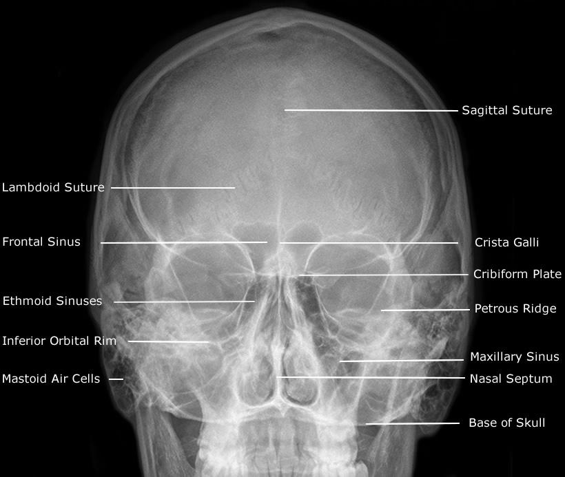
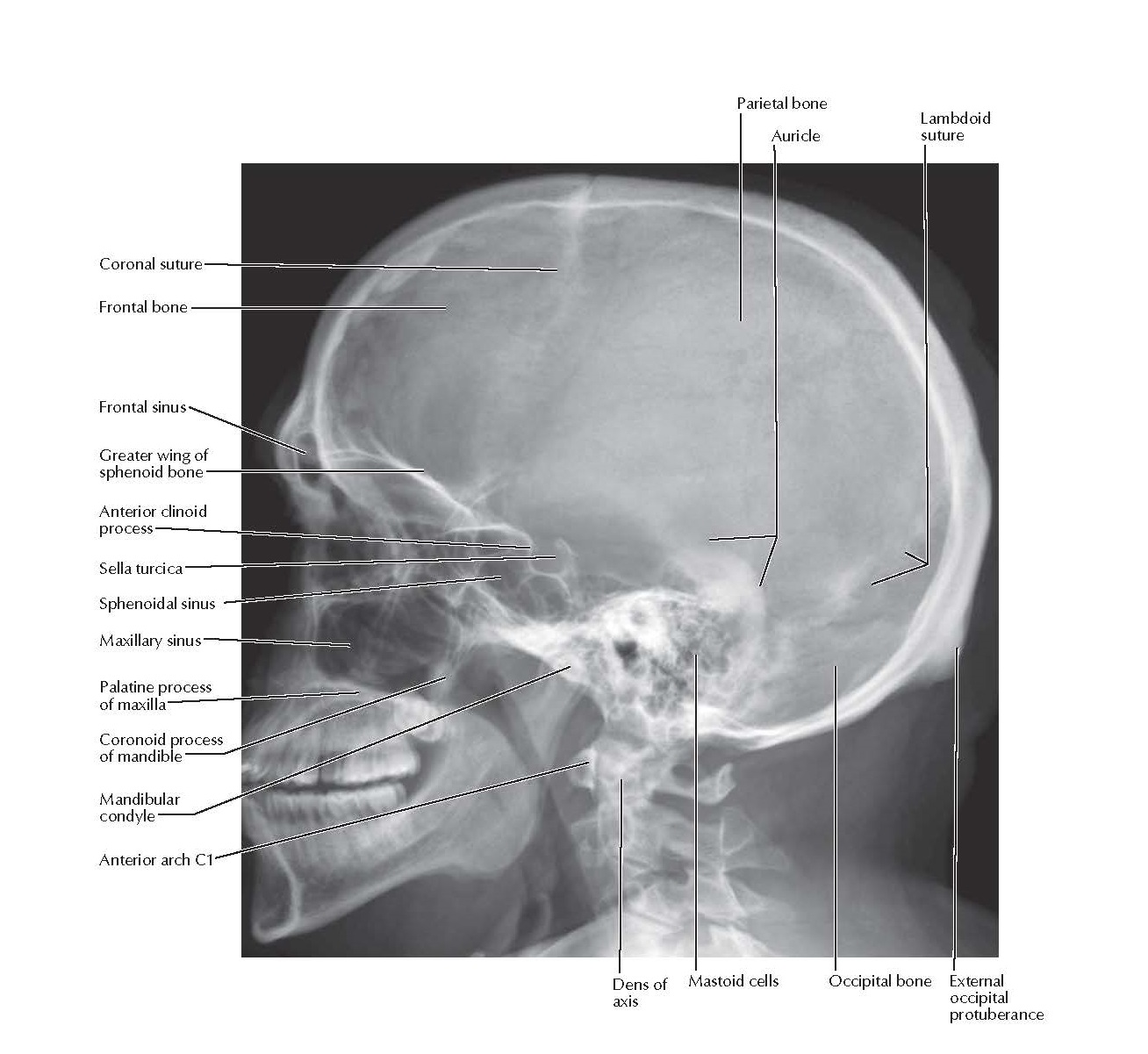
Skull Skull, a-p X-ray Sagittal suture Lambdoid suture Supra-orbital margin Lesser wing of sphenoid bone Hypophysial fossa Crista pyramidis (upper edge of petrous bone) Head of mandible Atlanto-occipital joint Lateral atlanto-axial joint Squama occipitalis Granular foveola Frontal sinus Jugum sphenoidale
Ap Skull X Ray Normal skull xray Image / Ap
Skull. The skull rests on the superior aspect of the vertebral column. It is composed of 22 separate bones divided into two distinct groups: 8 cranial bones and 14 facial bones. The cranial bones are divided further into the calvaria and floor ( Box 20-1 ). The cranial bones form a protective housing for the brain.
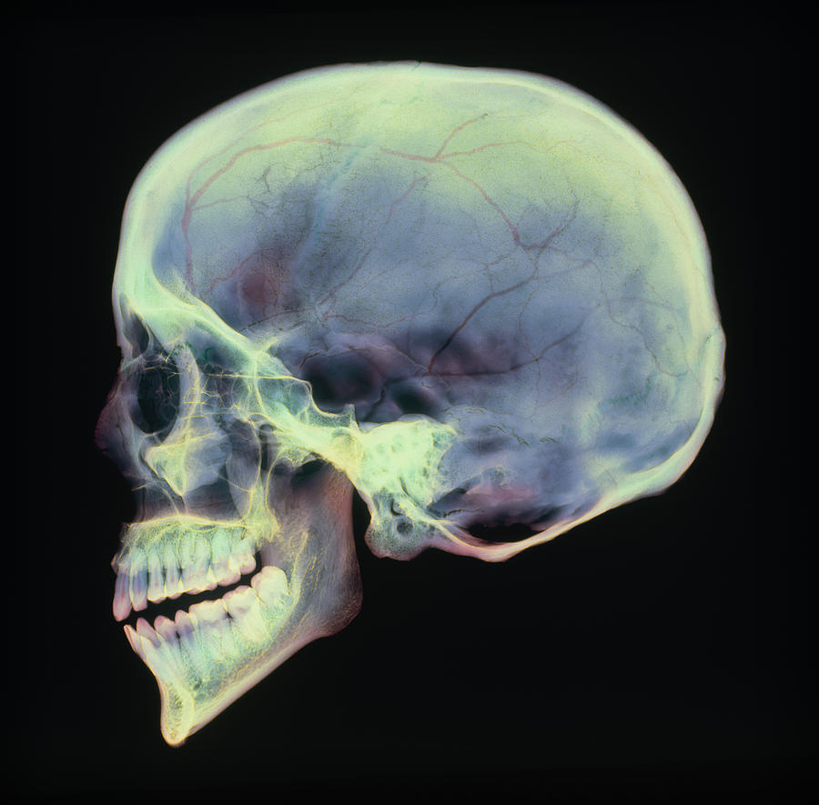
Human Skull, Xray Photograph by D. Roberts
The skull is a solid bony structure that encloses and protects the brain and other components of the central nervous system. It consists of 8 cranial bones and 14 facial bones (see our article on radiographic positioning of the face and mandible ). The back of the cranium consists of the occipital and right and left parietal bones.
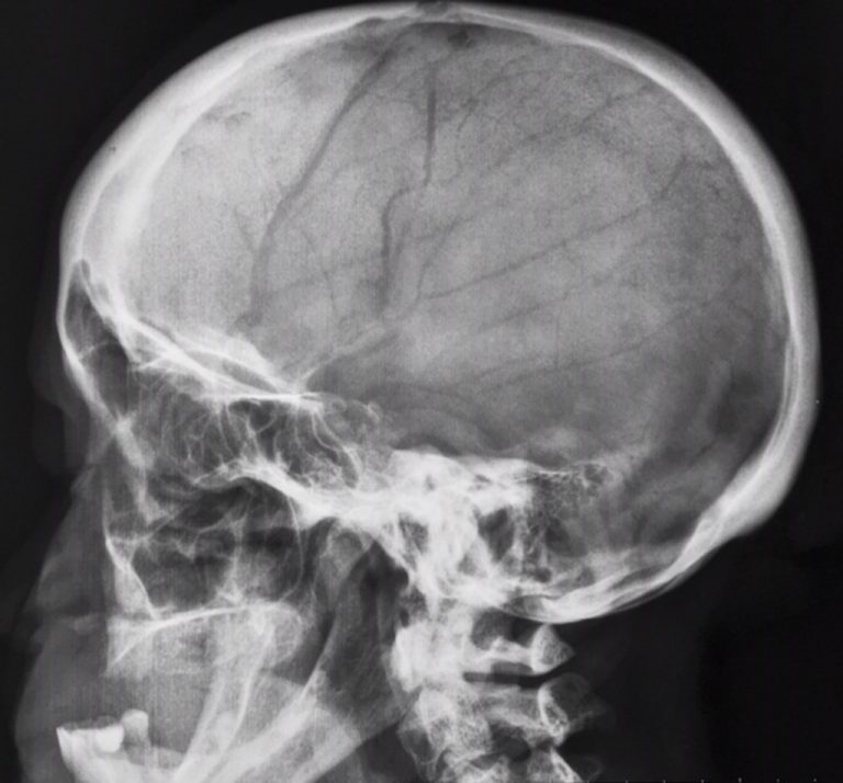
NORMAL LATERAL SKULLCHILD
A skull x-ray is a picture of the bones surrounding the brain, including the facial bones, the nose, and the sinuses. How the Test is Performed You lie on the x-ray table or sit in a chair. Your head may be placed in different positions. How to Prepare for the Test Tell the health care provider if you are pregnant or think you are pregnant.

Xray of skull ap and lateral view anatomy YouTube
Skull radiography is the radiological investigation of the skull vault and associated bony structures. Seldom requested in modern medicine, plain radiography of the skull is often the last resort in trauma imaging in the absence of a CT. Indications Skull radiographs are indicated for a variety of settings including: trauma

SKULL Caldwell Medical radiography, Radiology student, Radiology
The skull (TA: cranium) consists of 22 bones, excluding the three ossicles in each middle ear. All of the bones of the skull, except for the mandible, are connected to each other by sutures (fibrous joints) and are thus immobile. These 21 bones form the cranium, and are further subdivided into: neurocranium. calvaria; skull base (basicranium)
Skull xray
Abstract. In order to differentiate normal sutures and pathological fractures on radiological images, age-specific anatomy of the sutures should be understood. For the detailed visualization of the skull, a cadaver's sectioned images with real color and high resolution have been utilized. The sutures have been introduced using diverse imaging.

SKULL LATERAL VIEW
A skull X-ray is an imaging test doctors use to examine the bones of the skull, including the facial bones, the nose, and the sinuses. See a Body Map of the skull. It's an easy, quick, and.
Skull Lateral Radiograph Anatomy pediagenosis
Practical points the PA view decreases the radiation dose to the eyes compared with the AP view less magnification of the facial bones is achieved compared with the AP view overlap of facial bone structures makes it harder to evaluate the sinuses than with an angled view (e.g. Caldwell view) References Incoming Links
NORMAL LATERAL SKULL RADIOGRAPH
Skull X-ray What is a skull X-ray? X-rays use a small amount of radiation beams to make images. Standard X-rays are done for many reasons. They are done to diagnose tumors, infection, foreign bodies, or bone injuries. X-ray beams pass through body tissues onto treated plates. The more solid a structure is, the whiter it appears on the film.