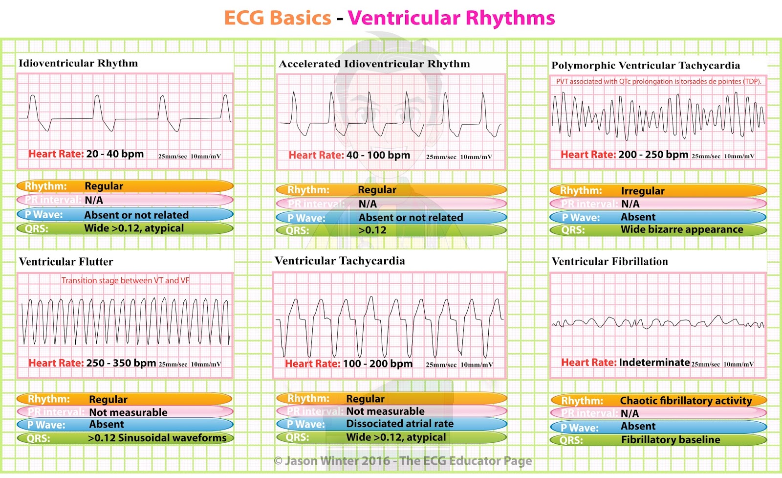
ECG Educator Blog Ventricular Rhythms
In some cases, patients may have an initial ECG without ST-segment elevation, making the diagnosis very challenging. 1 This is the case of STEMI equivalent patterns, such as hyperacute T waves, de Winter's pattern (dWp), Wellens syndrome, and posterior STEMI. 1 Among these, dWp is characterized by loss of R waves in the precordial leads.

De Winter T Wave • LITFL • ECG Library Diagnosis
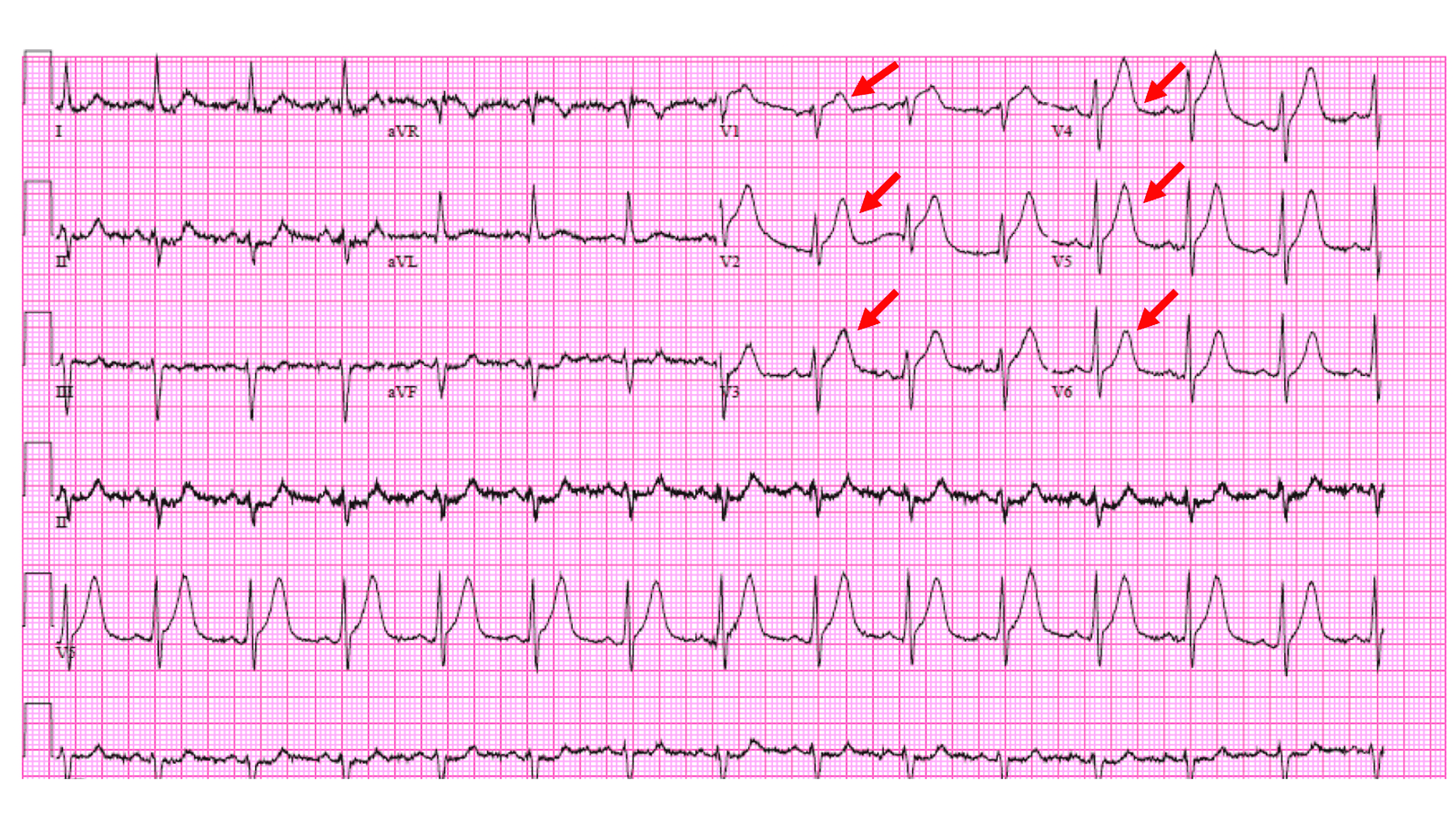
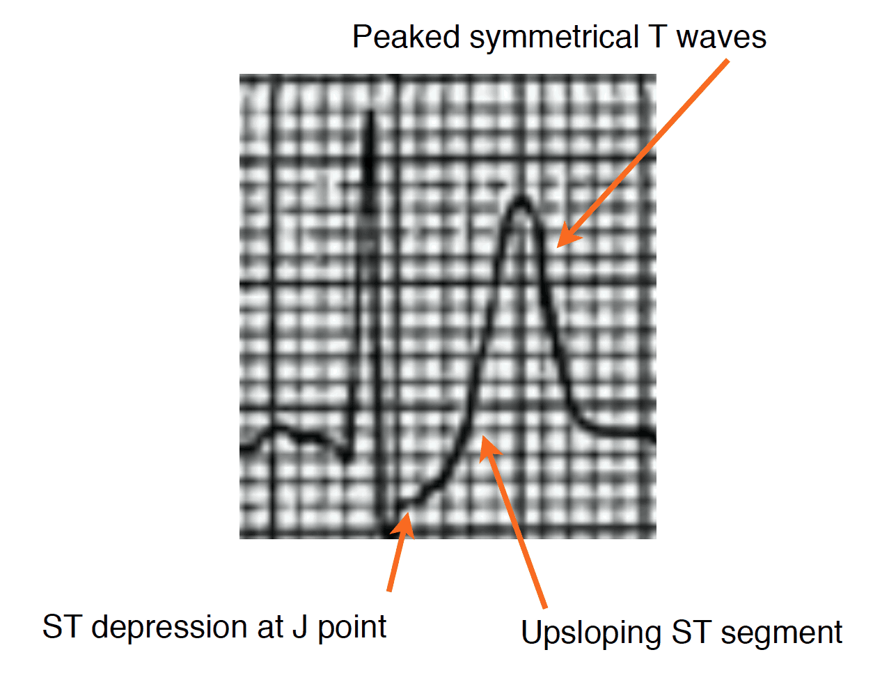
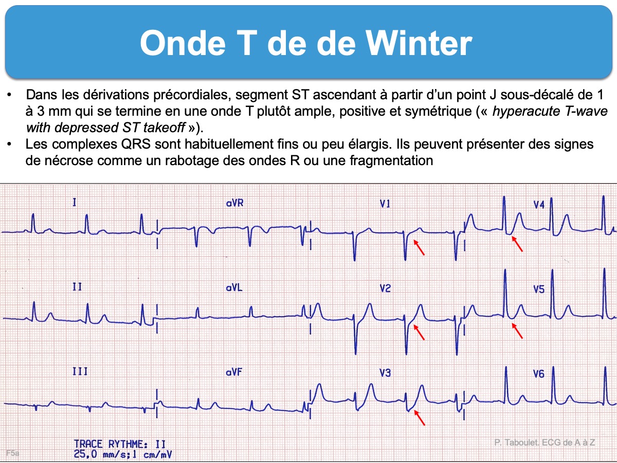
The main characteristics of the de Winter electrocardiogram (ECG) pattern are up-sloping ST-segment depression in the V 1 to V 6 leads, followed by tall and symmetrical T waves [ 1 ], which remain consistent with no evolutionary ECG changes.

ECG Basics Heart Blocks Diagnosis Cardiology MedStudent GrepMed
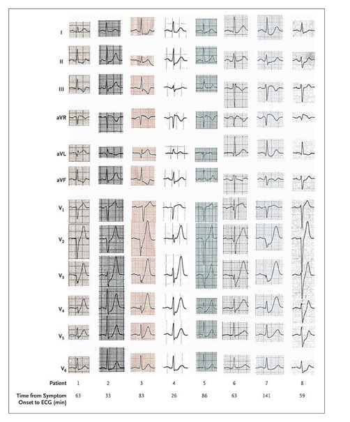
de Winter syndrome is a rare phenomenon, and it occurs in approximately 2% of patients with myocardial infarction. 2 It has a high predictive value for LAD occlusion. 4 Recent studies suggested that de Winter syndrome may be a transient event before progressing into typical STEMI ECG. 5 The exact electrophysiologic mechanisms involved are not.

ECG Educator Blog 05/16/16
De Winter's T waves 1 Background 3 References Background First identified in 2008 by Dr. DeWinter - characteristic pattern in 30 of his 1532-patient database of anterior MI [1] 2% of proximal LAD occlusions will have this presentation Represents an acute proximal occlusion (unlike Wellen's sign which represents a subacute process)

(PDF) Evolutionary de Winter pattern from de Winter ECG to STEMIA case report
First reported by Dutch Professor of Cardiology, Robbert J. de Winter in 2008, the de Winter ECG pattern is an anterior STEMI equivalent that presents without obvious ST segment elevation. These patients are suffering occlusion myocardial infarction (OMI) and require immediate reperfusion therapy. ECG Diagnostic Criteria
de winters ecg pattern blackclothesdigitalarttutorial
The De Winter ECG pattern has been reported to indicate acute left anterior descending coronary artery occlusion and is often considered to be an 'ST elevation myocardial infarction (STEMI) equivalent'.

ECG Rhythms De Winter's ST/T ECG changes in huge anterior wall MI
The De Winter T wave pattern is seen in approximately 2% of acute LAD occlusions and is often under-recognised by clinicians, leading to delays in reperfusion therapy. Most guidelines now consider this pattern a STEMI-equivalent and indication for immediate reperfusion therapy.. De Winter's T waves — ECG Diagnostic Criteria. Tall.
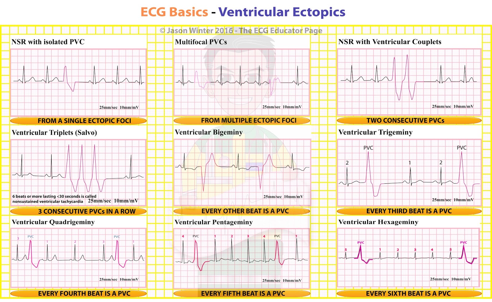
ECG Educator Blog Ventricular Ectopics
An electrocardiographic finding suggestive of impending myocardial infarction , the de Winter's pattern (or "de Winter's T-waves") describes an abnormality thought to be indicative of acute occlusion of the proximal left anterior descending coronary artery ( LAD) 2.
dual rhythm no murmur Victor Ogden
Whether the 'de Winter' ECG pattern is a static ECG pattern or a transient ischaemic phenomena preceding the evolution into an overt STEMI, it emphasizes the importance to realize serial ECGs when treating patients with an acute typical chest pain. In our patient, it is interesting to note that there were hyperacute T waves as an early sign.
Dewinter Ekg / de winter t dalgası anteriyor miyokard infarktüsü de quantum glassattery
We describe the case of a 56-year-old patient suffering from acute chest pain, presenting in our emergency department with a 'de Winter' ECG pattern: an upsloping ST-segment depression with tall symmetrical T waves associated with left anterior descending artery occlusion. Discussion
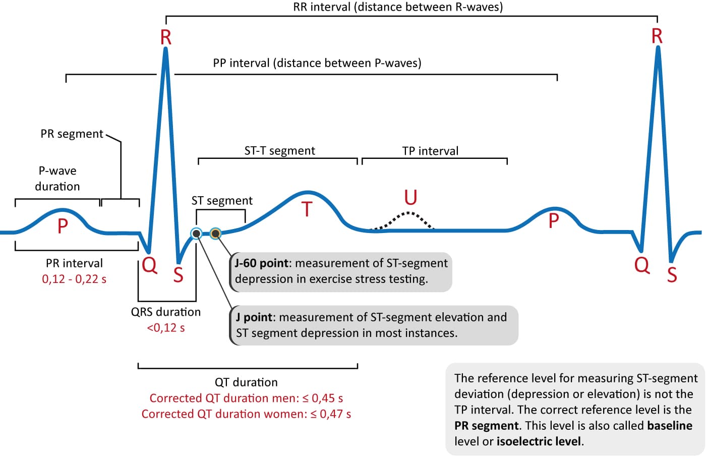
ECG interpretation Characteristics of the normal ECG (Pwave, QRS complex, ST segment, Twave
In 2008, de Winter et al described an ECG pattern suggesting that it should be considered an ST‐elevation myocardial infarction (STEMI) equivalent (de Winter, Verouden, Wellens, & Wilde, 2008 ), with the potential to predict critical stenosis or occlusion of the left anterior descending coronary artery (LAD). This ECG pattern typically.
ECG Rhythms De Winter's ST/T ECG changes in huge anterior wall MI
De Winter ST/T-Waves ECG abnormality described by de Winter et al. in 1998 Characterized by 1-3 mm of ST-depression with upright, symmetrical T-waves Changes are dynamic as you would expect with ACS (see Example 3 below) Suspicious for proximal occlusion of the LAD Represents approximately 2% of LAD occlusions

De Winter ST/TWaves ECG Medical Training Medical training, Medical, Medical examination
de Winter syndrome is a special equivalent of anterior ST-segment elevation myocardial infarction (STEMI) characterized by the absence of overt ST-elevation with upsloping ST-segment depression followed by tall symmetrical T-waves in the precordial leads, often associated with total occlusion of the proximal left anterior descending coronary art.
Dewinter Ekg Follow Up Ecg 75 Minutes After The Initial One Showing De Winter Download
Hence, hyperacute T-waves are the first ECG change in STE-ACS/STEMI. Since they are short-lived it is uncommon to encounter them in clinical practice. Recall that T-waves should not exceed 10 mm in chest leads and 5 mm in limb leads. de Winter's sign (persistent hyperacute T-wave syndrome) As mentioned above hyperacute T-waves have a short.

ECG showing typical de Winter's sign (syndrome) ECG learning
Winter is Coming Ed Burns Feb 2, 2022 Home LITFL Clinical Cases aka ECG Exigency 016 A 54-year old man presents by private vehicle to the Emergency Department with chest discomfort he described as " heartburn ." The pain is substernal and non-radiating. He is also mildly diaphoretic.

De Winter sign YouTube
Ischemic heart disease (IHD) is a major cause of morbidity and mortality. [1] Although there have been significant improvements in the overall management of patients suffering from acute coronary syndromes (ACS), this entity is still associated with a relevant clinical burden.